

56세/여 난소암 수술 후 3차 항암 이후 CT결과지 입니다.
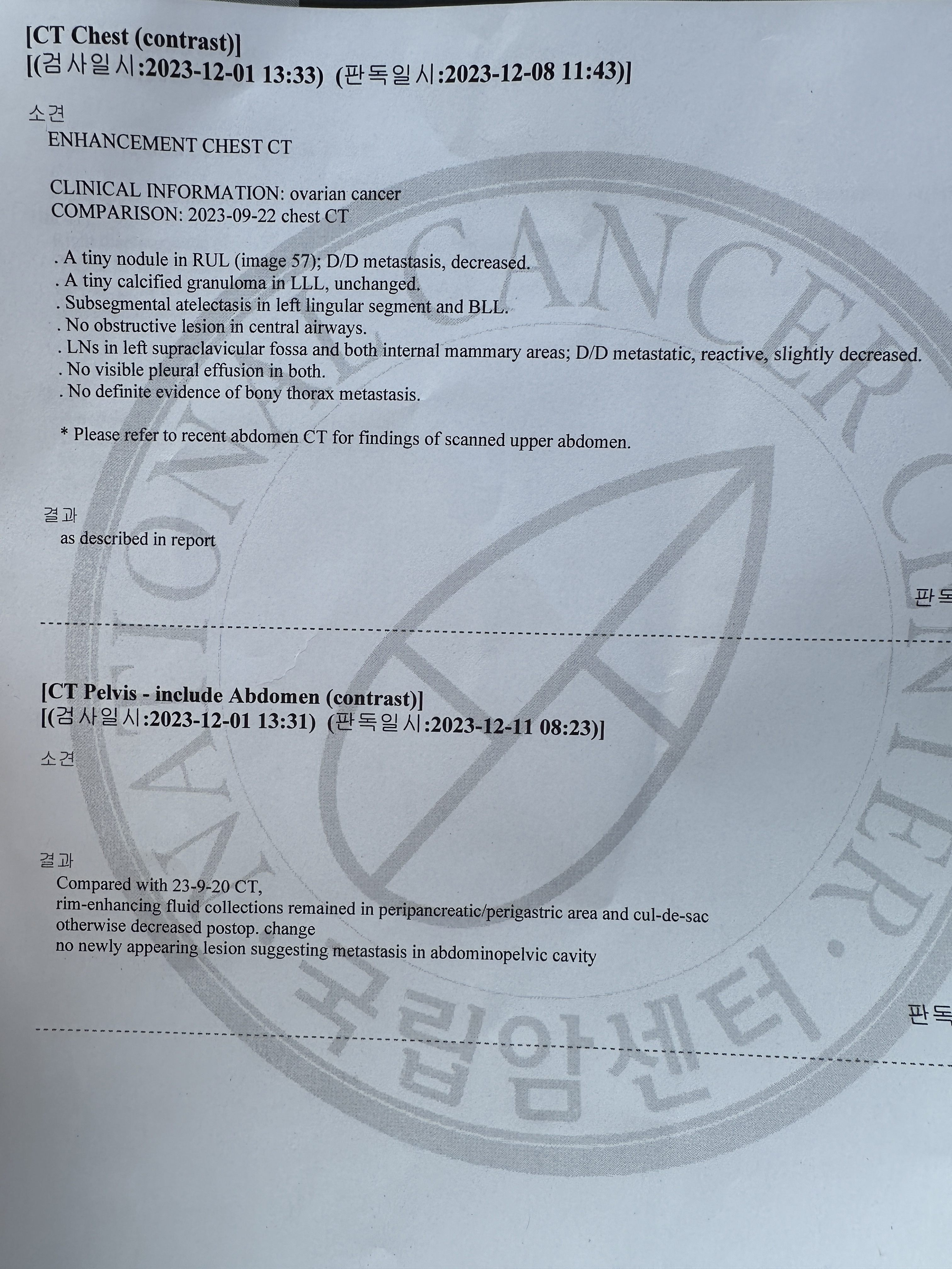
[CT Chest (contrast)]
[(검사일시:2023-12-01 13:33) (판독일시:2023120811:43)1
소견
ENHANCEMENT CHEST CT
CLINICAL INFORMATION: ovarian cancer
COMPARISON: 2023-09-22 chest CT
. A tiny nodule in RUL (image 57); D/D metastasis, decreased.
. A tiny calcified granuloma in LLL, unchanged.
. Subsegmental atelectasis in left lingular segment and BLL.
. No obstructive lesion in central airways.
. LNs in left supraclavicular fossa and both internal mammary areas; D/D metastatic, reactive, slightly decreased.
. No visible pleural effusion in both.
. No definite evidence of bony thorax metastasis.
* Please refer to recent abdomen CT for findings of scanned upper abdomen.
결과
as described in report
[CT Pelvis - include Abdomen (contrast)]
[(검사일시:2023-12-01 13:31) (판독일시:2023-12-11 08:23)]
소견
결과
Compared with 23-9-20 CT,
rim-enhancing fluid collections remained in peripancreatic/perigastric area and cul-de-sac otherwise decreased postop. change
no newly appearing lesion suggesting metastasis in abdominopelvic cavity