

※ 검사결과지를 보기 힘든 경우가 많습니다. ㅜㅌ
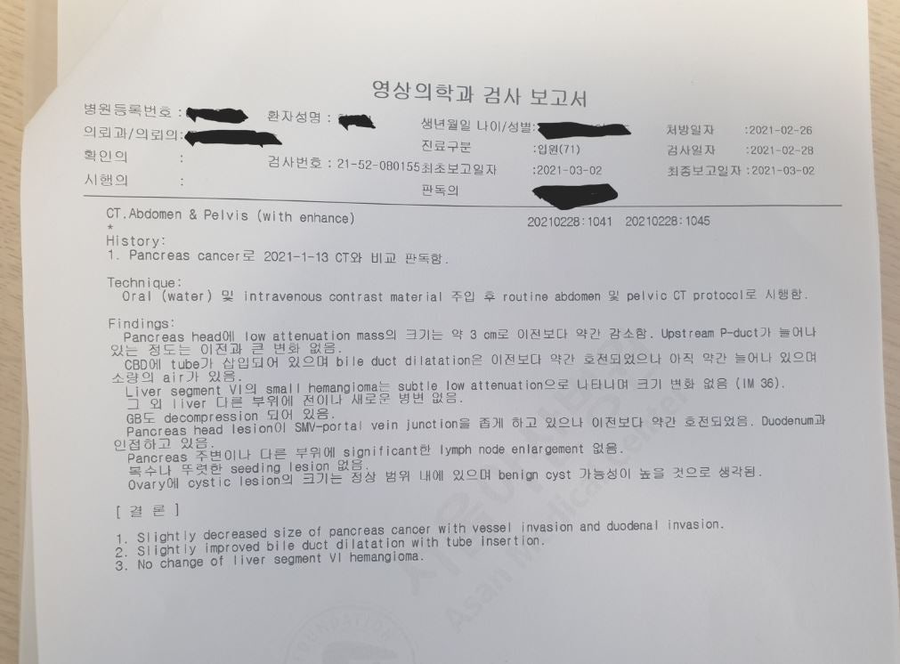
CT. Abdomen & Pelvis (with enhance)
20210228: 1041 202 10228: 1045
History:
1. Pancreas cancer로 2021-1-13 CT와 비교 판독함.
Technique :
Oral (water) 및 intravenous contrast material 수입 후 routine abdomen 및 pelvic CT protocol로 시행한.
Findings:
Pangreas head에 Tow attenuation mass의 크기는 약 3 cm로 이전보다 약간 감소함. Upstream P-duct가 늘어나있는 정도는 이전과 큰 변화 없음.
CBD에 tube가 삽입되어 있으며 bile duct dilatation은 이전보다 약간 호전되었으나 아직 약간 늘어나 있으며,
소량의 air가 있음.
Liver segment Vi의 small hemangiome는 subtle low atterustion 으로 니타나며 크기 변한 없음 (IM 36).
그 외 Tiver 다른 부위에 진이나 새로운 병변 없음.
GB 도 decompression 되어 있음.
Panoreas head lesionSMV-portal vein junctior 을 좁게 하고 있으나 이진보다. 약간 호전되었음. Duocenum과
인접하고 있음
Pancreas 주변이나 다른 부위에 significant한 lymph node en largement 없음. .
복수나 뚜렷한 seeding lesion 없음.
Ovary에 cystic lesion의 크기는 정상 범위 내에 있으며 benign cyst 가능성이 높을 것으로 생각됨.
[결론]
1. Slightly decreased size of pancreas cancer with vessel invasion and duodenal invasion.
.
2. Slightly improved bile duct dilatation with tube insertion
3. No change of liver segment VI hemangioma.
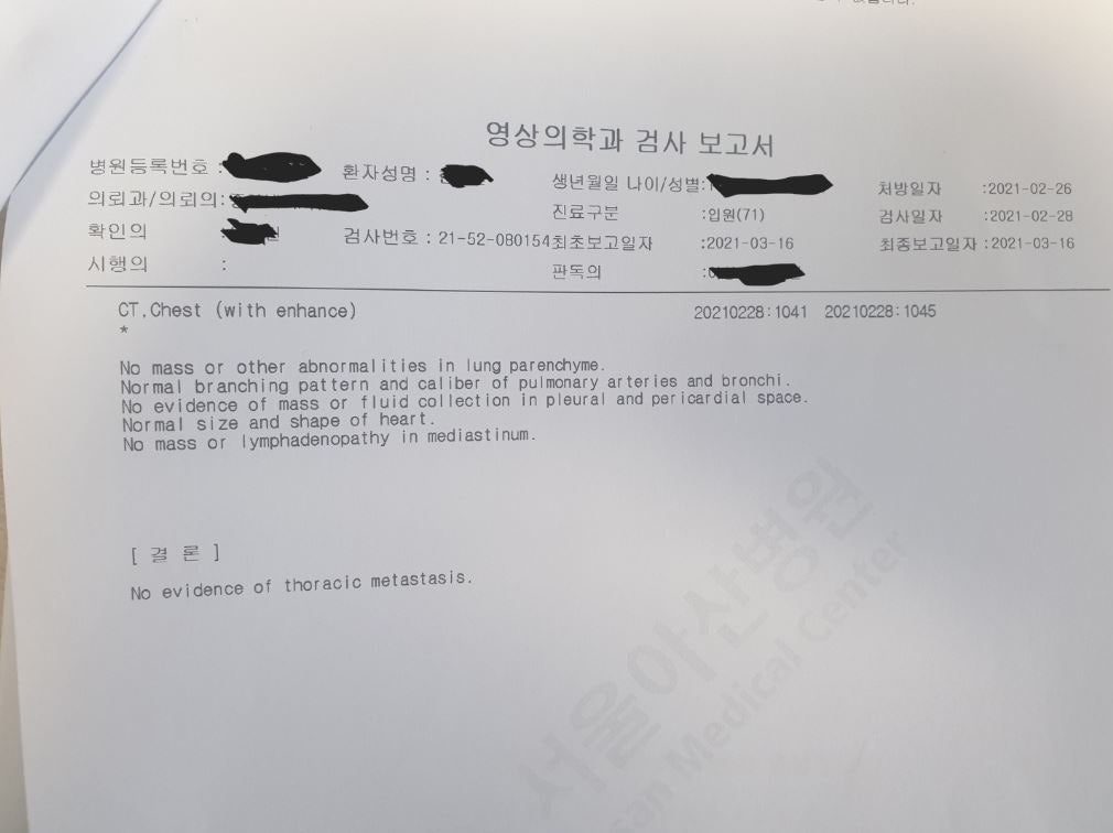
CT. Chest (with enhance)
20210228: 1041 202 10228: 1045
#
No mass or other abnormalities in lung parenchyme.
Normal branching pattern and callber of pulmonary arteries and bronchi
No evidence of mass or fluid collection in pleural and pericardial space
Normal size and shape of heart.
No mass or lymphadenopathy in mediastinum.
[결론]
No evidence of thoracic metastasis.